In Fricktal, a landscape in the north of Switzerland, a large number of skeletons of herbivorous dinosaurs have been found in recent years. These plateosaurus lived about 200 million years ago and reached a body length of 5 to 8 meters. Of the approximately 80 (partial) skeletons found so far, only about 10% were completely preserved with skulls. It was all the more special for us to have such a skull in the studio (on loan from the Sauriermuseum Frick).
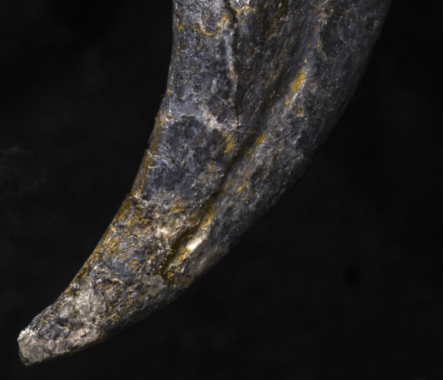
During the one-day shooting session, it was important to take beautiful pictures on the one hand and to create very high-resolution and detailed models of the surface structures on the other hand by means of RTI. In the case of the skull, the main interest was the dentition and its wear marks; in the case of the claw (more precisely, the bone core of one), the focus was on an overall shot and a detail of the (blood) supply channel at the tip.
To get to know our model, we first took care of the conventional overall image of the skull.

For orientation: The skull was damaged from above by an excavator and is now visible from the underside. Therefore, the teeth on the lower left of the image correspond to the upper right jaw.
What appears to be a fracture on the lower left is probably the boundary between two skull bones. Dinosaur skulls consist of about 60 small bone parts.
Because the main interest was on the teeth, we first staged them perfectly - two Picolites were used for this: For the front (upper) part, a narrow honeycomb grid was precise enough; for the back teeth further away from the light, a Projection attachment (slightly out of focus) was the best choice, as its light cone could be perfectly adjusted even over the greater distance. The flat light angle of these two light sources perfectly highlighted the surface structure.
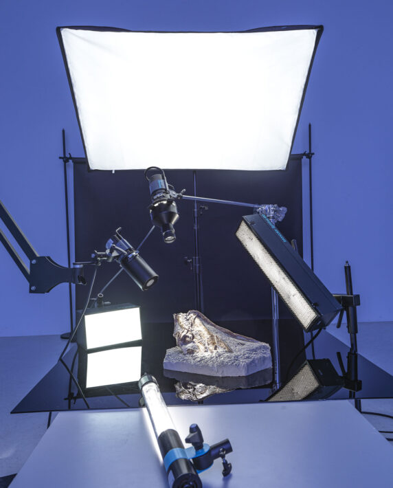
The other lamps were also positioned (as backlights) to emphasize the three-dimensionality of the skull. A Striplite 60 (with honeycomb) was used behind the subject on the right, and a Picobox was placed directly on the shooting table behind the skull on the left.
After setting up these four lamps, it was still a matter of achieving a discreet brightening of the previously very dark shadows. To do this, a large softbox was placed over the skull (and secured several times to guarantee it wouldn't fall off and damage the subject). Because even this brightening did not come from the direction of the camera (which would have made everything look much flatter) a final, precise light was needed to brighten only the front edges of the base. For this purpose, a Litestick was used.
For this total shot, I decided on a lighting setup that seemed sensible to me. But it is quite conceivable that for scientific purposes not "the most beautiful" light angle is useful, but just another one. The virtual illumination of a 2-dimensional RTI surface model of the 3-dimensional surface can be adjusted afterwards. This means that you do not have to decide on a final lighting situation when taking the picture but can change it at any time as required.
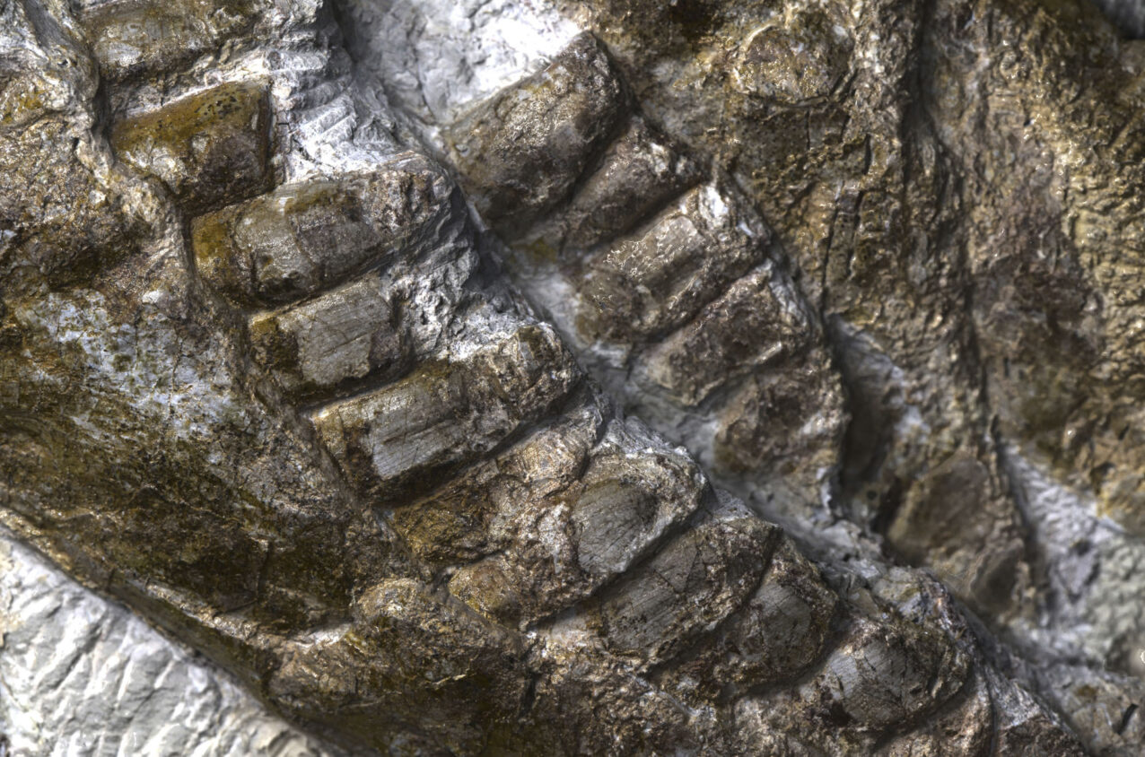
This detailed image of the dentition was taken with a broncolor Scope D50 in the visible light spectrum (Scope D50 also allows images in the UV and IR range).
The AUTHENTICA software that completes the Scope D50 also allows not only the illumination, but even the materiality of objects to be changed virtually. In the following example (based on the same surface model), true colors were omitted and the gloss level was increased instead. In combination with a flat, hard, virtual light, previously hidden properties of the surface became visible:
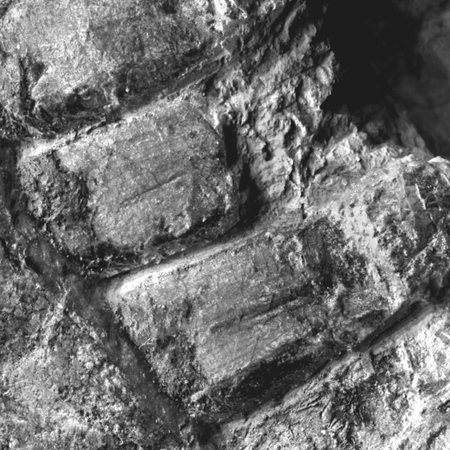
The layered structure of the teeth is still recognizable. In contrast, the upper edges of the teeth, which are otherwise finely toothed, are worn or not preserved in this case.
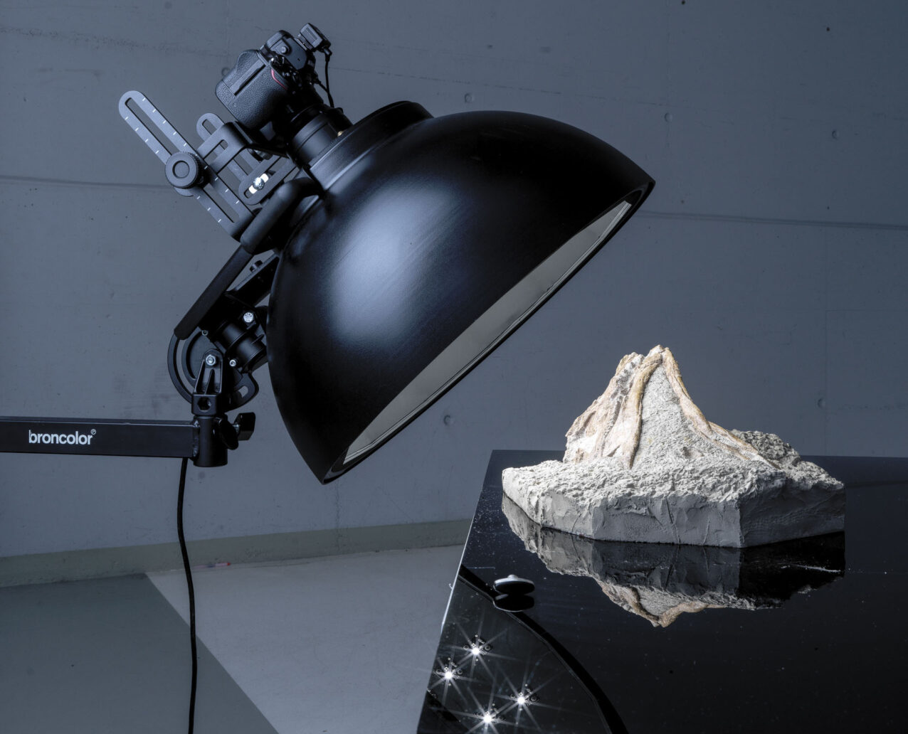
In the case of the bone core of the claw, too, the aim was not "only" to create a beautiful photograph, but also to obtain as much three-dimensional information as possible.
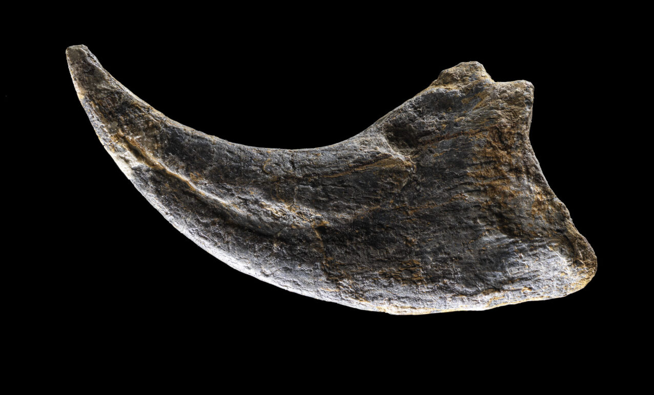
Conventional photography therefore exclusively used hard (narrow) light shapers in a very flat light angle: a Striplite 60 in an angle of almost 90 degrees and a Litestick even in a light backlight. The "brightening" from the other side was also hard, as a mirror reflected only the hard main lights.
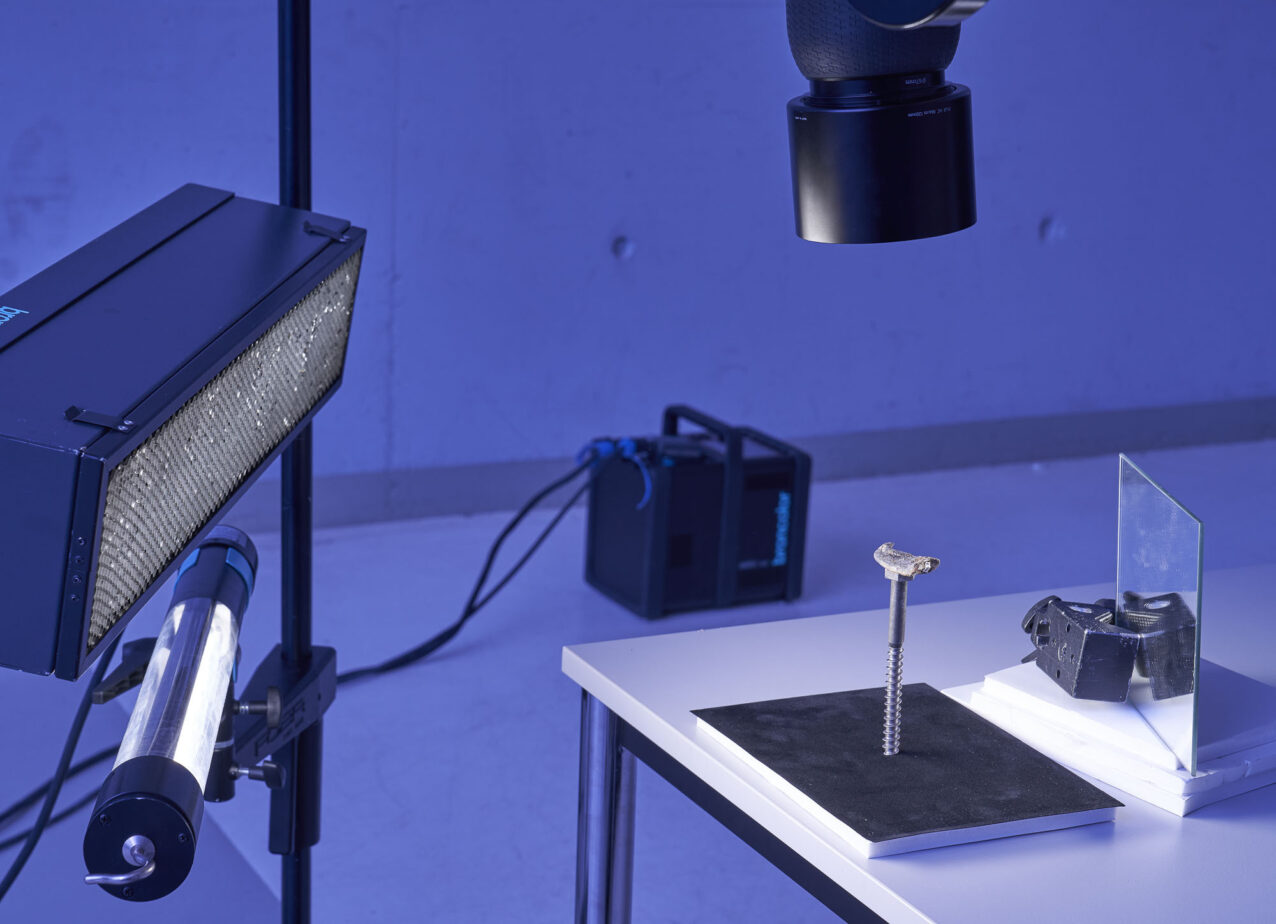
As described in the introduction, the main scientific interest is in the blood supply channel that lies between the bone core photographed here and the coating of keratin (not preserved), which supplied blood and oxygen to the periosteum there. The RTI model was again made with a Scope D50 in visible light and the surface slightly shinier (but in original colors).
Use your mouse or finger to interact with the below surface model
Enter in the full-screen mode using the square button, zoom. Click, hold and drag your mouse to change the angle of the light on the surface model.